


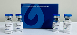


CHO-K1/M5 Stable Cell Line
Muscarinic acetylcholine receptors belong to a superfamily of seven-TM-domain receptors that interact with G-proteins to initiate intracellular responses. Five muscarinic receptor subtypes have been identified, named M1 through M5. Receptors of the M5 receptor subtype couple through the Gq/11 class of G-proteins and activate the phospholipase C pathway. Activation of this pathway in turn leads to increases in free intracellular calcium levels as inositol triphosphate mediates release of calcium from the endoplasmic reticulum. RT-PCR reveals that M5 mRNA is quite uniformly expressed in brain. However, there is little data regarding the expression and function of the M5 receptor in peripheral tissues. Currently, it is clear that the M5 receptor, due to the high likelihood that its distribution is restricted to the CNS, probably plays a discrete role in dopaminergic transmission. Although the identification of M5 expression in salivary glands and iris-ciliary muscle suggests a broader role, the data on this is sparse and requires extensive confirmation.
| 询价 | |
| M00186 | |
|
|
|
|
|
|
|
|
|
| 咨询产品问题 | |