| Products/Services Used | Details | Operation |
|---|---|---|
| Gene Synthesis | The cDNA sequence encoding the murine plasma kallikrein protease domain, from I16 to A255, was synthesized (GenScript, USA) introducing C122S mutation, and the Kex2 sites following Xho I and Sal I restriction sites were at the 5’ and 3’ ends. The DNA fragment was inserted into the pPICZαA vector. The recombinational plasmid containing mPK was linearized using Sac I and transformed into the Pichia pastoris X-33 strain through electronic transformation (1500 V, 25 μF, 200 Ω, 7.2 ms) with JY 2000-1B gene transfection apparatus (Scientz, NingBo, China). After incubation in 1 M sorbitol for 2 h at 30 °C, the single colony was isolated from an YPD plate (1% yeast extract, 2% peptone, 2% glucose, 2% agar) containing 100 μg/mL Zeocin. The colony was then cultured in 2 mL BMGY (1% yeast extract, 2% peptone, 100 mM potassium phosphate, pH 6.0, 1.34% yeast nitrogen base, 4 × 10⁻⁵ % biotin, 1% glycerol) at 30 o C to an OD600 reading of 2~6, and sequentially cultivated in 8 mL BMMY (1% yeast extract, 2% peptone, 100 mM potassium phosphate, pH 6.0, 1.34% yeast nitrogen base, 4 × 10⁻⁵ % biotin, 1% methanol) for up to 120 h at 28 °C with shaking at 220 rpm. | Get A Quote |
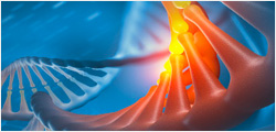
Plasma kallikrein (PK), a serine protease in the trypsin clan (SA), plays critical roles in many physiological and pathological pathways. Regulating the abnormal activity of PK has been successfully used in the clinical therapy of hereditary angioedema. In this study, the serine protease domain of murine plasma kallikrein (mPK) was expressed in the pichia pastoris system. The recombinant protein was a glycosylated active enzyme after purification by the cation exchange and size-exclusion chromatography, and was crystallized at the precipitant condition of 25% PEG 3350, 0.1 M Tris-HCl pH 8.5 and 0.1 M NaCl. The crystal structure of mPK was determined at 2.6 Å. This is the first published crystal structure of mP... More