首页
-
试剂服务
-
产品
-
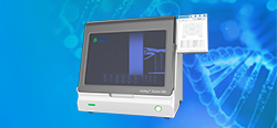
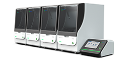
- AmMag™ Quatro 1300全自动蛋白/抗体磁珠纯化系统New!
- CytoSinct™ 1000 细胞分选仪 & 管路Hot!
- AmMag™ Quatro 1100全自动小型磁珠纯化系统Hot!
- AmMag™ Quatro 1400全自动多功能磁珠纯化系统
- eBlot™ 快速湿转仪Hot!
- eStain® L1蛋白染色仪
- eZwest™ 全自动蛋白印迹系统
- GenBox™ 电泳槽
- 电泳仪电源
- AmMag™ Quatro 1400 全自动多功能磁珠纯化系统Hot!
- AmMag™ Quatro 1100 全自动多功能小型磁珠纯化系统Hot!
- AmMag™ Quatro 1300 全自动蛋白/抗体磁珠纯化系统New!
- 亲和纯化磁珠
- 内毒素检测与去除
- AmMag™ Quatro 1300全自动蛋白/抗体磁珠纯化系统New!
- StrepCaptureXP高纯度蛋白纯化工具Hot!
- 亲和层析介质
- 亲和纯化磁珠
- His标签蛋白纯化
- 抗体纯化
- 内毒素去除&检测
- DYKDDDDK标签蛋白纯化Hot!
- 耐碱Protien A 磁珠
- SurePAGE™ 蛋白预制胶Hot!
- YoungPAGE™ 蛋白预制胶New!
- eBlot™ 快速湿转仪Hot!
- eStain® L1蛋白染色仪Hot!
- eZwest™ 全自动蛋白印迹系统
- 蛋白Marker
- GenBox™ 电泳槽
- 电泳仪电源New!
- GMP级别细胞分选和激活Hot!
- CytoSinct™ 细胞分选磁珠
- Enceed™ 细胞激活试剂Hot!
- CytoSinct™ 分选柱 & 磁力架Hot!
- CytoSinct™ 1000 细胞分选仪 & 管路
- 纳米磁珠定制化平台New!
- 标签抗体Hot!
- Anti-VHH AntibodyHot!
- Anti-scFv AntibodyHot!
- 抗独特型抗体Hot!
- Anti-AAV抗体
- Anti-CD抗体
- Anti-Payload抗体Hot!
- 一抗
- 二抗
- 药物代谢试剂盒
- 病毒载体质控试剂盒
- -- 慢病毒滴度p24试剂盒Hot!
- -- MuLV滴度p30检测试剂盒New!
- -- AAV滴度衣壳蛋白ELISA试剂盒
- 工艺残留质控试剂盒
- -- T7 RNA Polymerase ELISA试剂盒New!
- -- BSA ELISA试剂盒
- -- Cas9 ELISA试剂盒New!
- -- 蛋白A ELISA试剂盒New!
- -- His标签蛋白检测&纯化
- -- dsRNA ELISA试剂盒New!
- 应用领域
-
活动&资源
- 关于我们
- 登录 用户中心 退出
- 注册
