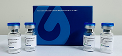
Product Description | Recombinant CHO-K1 cells stably overexpress human neuropeptide S receptor 1 (NPSR1) isoform B on the surface and contain high levels of G protein Gαs and Gαq to couple with the receptor in downstream signaling pathways. |
Culture Properties | Adherent. |
Stability | Stable through more than 16 passages with no significant changes in assay performance or expression profile. |
Size | Two vials of frozen cells (>1×10⁶ per vial in 1 mL). |
Storage | Store cells in liquid nitrogen immediately upon receipt. Thaw and recover cells within one year from the date received. |
Culture Medium | Ham’s F-12K (Kaighn’s), 10% FBS, 200 μg/ml Zeocin (Cat. No. R250-01, Life Technologies), 100 μg/ml Hygromycin B (Cat. No. 10687010, Life Technologies) |
Complete Growth Medium | Ham’s F-12K (Kaighn’s), 10% FBS |
Freeze Medium-DATA | 45% Ham’s F-12K (Kaighn’s) (Cat. No. 21127, Life Technologies), 45% FBS (Cat. No. 10099-14, Life Technologies), 10% DMSO (Cat. No. D2650, Sigma) |
Figure 1. Neuropeptide S-induced concentration-dependent stimulation of intracellular calcium mobilization in CHO-K1/NPS1b/Gα15 cells. The cells were loaded with Calcium-4 prior to being stimulated with agonist neuropeptide S. The intracellular calcium change was measured by FLIPR. The relative fluorescent units (RFU) were normalized and plotted against the log of the cumulative doses of neuropeptide S (Mean ± SEM, n = 3). The EC50 of neuropeptide S on CHO-K1/NPS1b/Gα15 cell was 0.20 μM.
Notes:
EC50 value is calculated with four parameter logistic equation:
Y=Bottom + (Top-Bottom) / (1+10^((LogEC50-X)*Hill Slope))
X is the logarithm of concentration. Y is the response
Y is RFU and starts at Bottom and goes to Top with a sigmoid shape.
»
Figure 2. Dose dependent stimulation of intracellular IP-One accumulation upon treatment with neuropeptide S on CHO-K1/NPS1b/Gα15 cells. d2 acceptor fluorophore-labeled IP-One (Cat. No. 62IPAPEB; Revvity) and intracellular IP-One in CHO-K1/NPS1b/Gα15 cells competitively bind with Europium Cryptate-labeled anti-IP-One antibody. The FRET signal decreases as the intracellular IP-One concentration rises and was measured by plate reader (Pherastar, BMG). The EC50 of neuropeptide S on CHO-K1/NPS1b/Gα15 cells was 0.10 μM. »
For research use only. Not intended for human or animal clinical trials, therapeutic or diagnostic use.