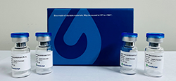
Product Description | Recombinant CHO-K1 cells stably overexpress human adenosine A1 receptor (ADORA1) on the surface and contain high levels of G protein Gαi to couple with the receptor in downstream signaling pathways. |
Culture Properties | Adherent |
Stability | Stable through more than 16 passages with no significant changes in assay performance or expression profile. |
Size | Two vials of frozen cells (>1×10⁶ per vial in 1 mL) |
Storage | Store cells in liquid nitrogen immediately upon receipt. Thaw and recover cells within one year from the date received. |
Culture Medium | Ham’s F-12K (Kaighn’s), 10% FBS, 200 μg/ml Zeocin (Cat. No. R250-01, Life Technologies), 200 μg/ml Hygromycin B (Cat. No. 10687010, Life Technologies) |
Complete Growth Medium | Ham’s F-12K (Kaighn’s), 10% FBS |
Freeze Medium-DATA | 45% Ham’s F-12K (Kaighn’s) (Cat. No. 21127, Life Technologies), 45% FBS (Cat. No. 10099-141, Life Technologies), 10% DMSO (Cat. No. D2650, Sigma) |
Figure 1. NECA-induced concentration-dependent stimulation of intracellular calcium mobilization in CHO-K1/ADORA1/Gα15 cells. The cells were loaded with Calcium-4 (Cat. No. R8142; Molecular Devices) prior to stimulation with ADORA1 agonist, NECA (Cat. No. HY-103173, MCE). The intracellular calcium change was measured by FLIPRTETRA. The relative fluorescent units (RFU) were recorded and plotted against the log of the cumulative doses of NECA (mean ± SEM, n = 3). The EC50 of NECA on CHO-K1/ADORA1/Gα15 cells was 0.12 μM.
Notes:
EC50 value is calculated with four parameter logistic equation:
Y=Bottom + (Top-Bottom) / (1+10^ ((LogEC50-X)*Hill Slope))
X is the logarithm of concentration. Y is the response.
Y is ∆RFU and starts at Bottom and goes to Top along a sigmoid curve.
»
Figure 2. Dose dependent increase of intracellular cAMP accumulation upon treatment with CGS-15943 in CHO-K1/ADORA1/Gα15 cells. d2 acceptor fluorophore-labeled cAMP (Cat. No. 62AM4PEC; Revvity) and intracellular cAMP in CHO-K1/ADORA1/Gα15 cells competitively bind with Europium Cryptate-labeled anti-cAMP monoclonal antibody. The FRET signal decreases as the intracellular cAMP concentration rises and was measured by plate reader (Varioskan, Thermo Fisher). The EC50 of CGS-15943 on CHO-K1/ADORA1/Gα15 cells was 67.83 nM. »
For research use only. Not intended for human or animal clinical trials, therapeutic or diagnostic use.