Specificity |
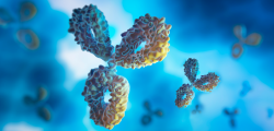
GenScript’s THE™ PEG Antibody, mAb, Mouse has high affinity for the PEG
backbone. The antibody binds to the variety of PEG such as PEG40K, PEG20K,
PEG5K, PEG12, PEGylated drugs and PEG conjugates. |
Host Species | Mouse |
Immunogen | PEG conjugated to KLH |
Conjugate | Unconjugated |
Working concentrations for specific applications should be empirically determined by the investigator. The appropriate concentrations may be affected by secondary antibody affinity, antigen concentration, the sensitivity of the detection methods, temperature, the length of the incubations, and other factors. The suitability of this antibody for applications other than those listed below has not been determined. | |||||||||||
|
Form | Lyophilized |
Storage Buffer |
Lyophilized with PBS, pH 7.4, containing 0.02% sodium azide. |
Reconstitution |
Reconstitute the lyophilized powder with deionized water (or equivalent)
to a final concentration of 0.5 mg/mL. For long-term storage, adding glycerol
to a final concentration of 50% (v/v) in the reconstituted antibody solution is
recommended. |
Storage Instructions |
The lyophilized product remains stable for up to 1 year at -20°C from the
date of receipt. The reconstituted antibody can be stored for 2-3 weeks at
2-8°C or for up to 12 months at -20°C or below. Avoid repeated freezing and
thawing cycles. |
Purification | Affinity chromatography |
Isotype | Mouse IgM |
Clonality | Monoclonal |
Clone ID | 5E10E9 |
Note |
Some
reagents such as Tween-20, Triton X-100 and NP-40 interfere with the PEG
antibody interactions due to their structural similarity to PEG. Antibody
solution should not contain such reagents in the assay of PEGylated molecules. GenScript can customize this product per customer's request including product size, buffer components, etc. |
Figure 6. Sensitivity comparison of THE™ PEG Antibody, mAb, Mouse (GenScript, A01795; blue), and competitor’s Mouse Anti-PEG mAb (Company A, Clone AGP4; red) by indirect ELISA. The test results show that GenScript’s PEG Antibody could detect PEG with different molecular weights and has better reactivity to PEG40K, PEG20K, PEG5K and PEG12 (Pierce, MES(PEG)12) than the competitor’s product. »
Figure 7. Sensitivity comparison of THE™ PEG Antibody, mAb, Mouse (GenScript, A01795; red) and competitor’s Mouse Anti-PEG mAb (Company A, Clone AGP4; yellow) by indirect ELISA. The test results show that GenScript’s PEG Antibody had better reactivity to PEG20K than the competitor’s product. »
Figure 1. Double-antigen sandwich ELISA analysis of THE™ PEG Antibody, mAb, Mouse (GenScript, A01795). PEG antibody was captured by PEG40K-OVA and detected by PEG20K-Biotin and Streptavidin-HRP conjugate. The test result shows that GenScript’s PEG Antibody is suitable for Bridging ELISA application. »
Figure 5. Sensitivity comparison of THE™ PEG Antibody, mAb, Mouse (GenScript, A01795; red), and competitor’s Mouse Anti-PEG mAb (Company A, Clone AGP4; black) by indirect ELISA, with plate coated with PEG40K-OVA. The test results show that GenScript’s PEG Antibody had better reactivity to PEG40K-OVA than the competitor’s product. »
Figure 8. The half maximal inhibitory concentration (IC50) comparison of THE™ PEG Antibody, mAb, Mouse (GenScript, A01795; red) and competitor’s Mouse Anti-PEG mAb (Company A, Clone AGP4; black) in competitive ELISA. The ELISA plate was coated with PEG40K-OVA, and unconjugated PEG40K was used for determining the IC50 of anti-PEG mAb. The test results showed that GenScript’s PEG Antibody had lower IC50, therefore, better specificity and higher affinity to PEG were validated. »
Figure 4. Sensitivity comparison of competitor’s Mouse Anti-PEG mAb (Company A, Clone AGP4 ) with THE™ PEG Antibody, mAb, Mouse (GenScript, A01795) by Western Blot.
A,C: PEGylated drug (PEGASYS®, Peginterferon alfa-2a)
B,D: Interferon Alfa 2A protein
In both panels A and B, the primary antibody of was Mouse Anti-PEG mAb (Company A, Clone AGP4) used at 0.2 µg/mL concentration;
In both panels C and D, the primary antibody of was THE™ PEG Antibody, mAb, Mouse (GenScript, A01795) used at 0.2 µg/mL concentration;
The test results show that GenScript’s PEG Antibody has better sensitivity and specificity than the competitor’s product.
»
Figure 2. Immunohistochemistry analysis of mouse kidney tissue (Paraffin embedded) using THE™ PEG Antibody, mAb, Mouse (GenScript, A01795; 10 µg/mL) after mice were injected with BSA-PEG as an immunogen. The test results show that GenScript’s PEG Antibody is suitable for IHC application. »
Figure 3. In-Cell ELISA analysis of PEGylated drug-HepG2 interaction using THE™ PEG Antibody, mAb, Mouse (GenScript, A01795; 1 µg/mL) after HepG2 cells were treated with PEGylated drug (PEGASYS®, Peginterferon alfa-2a) at different concentrations. The test results show that GenScript’s PEG Antibody is suitable for the analysis of PEGylated drug-cell interaction. »
Target Background |
PEG (Polyethylene glycol) is a polyether compound with many applications
from industrial manufacturing to medicine. PEGylation is a technology that
covalently couples non-toxic, hydrophilic polyethylene glycol (PEG) to the
drug. It is an FDA-approved method for the delivery of protein drugs. PEG
modification can reduce the drug immunogenicity and antigenicity. PEGylated
drug decelerates renal excretion, improves stability towards proteolysis and
increases its half-life in blood. Accurate and sensitive quantification of PEG
conjugates is important for PEG conjugated product development and
pharmaceutical study. Polyethylene glycol (PEG) antibody is a useful tool for
the detection of PEGylated molecules. |
Synonyms |
Anti-Polyethylene glycol antibody, mAb, Mouse |
For laboratory research use only. Direct human use, including taking orally and injection and clinical use are forbidden.