


GFP Antibody, pAb, Rabbit
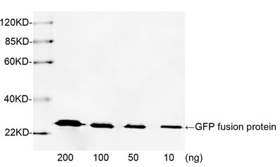
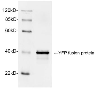
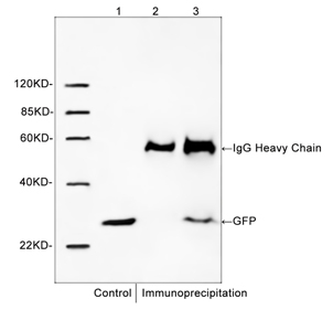
GenScript Rabbit Anti-GFP Polyclonal Antibody reacts with GFP fusion proteins. The antibody also reacts with other variants of GFP, such as CFP, YFP, eGFP and GFPuv.
| ¥990 在线订购包邮(港澳台除外) | |
| A01388-40 | |
|
|
|
|
|
|
|
|
|
| 咨询产品问题 | |